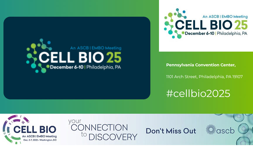
- I. Introduction: The Wind Rises from the "Bell Tower"—The Site Selection and Historical Context of Cell Bio 2025, the Premier Bio US Conference
- II. Interpreting the "Heat Map": Three Emerging "Core Battlefields" at Cell Bio 2025 – The Leading Bio US Conference
- III. Approaching the "Singularity": The "X Factor" Technologies Driving the Next Leap at Cell Bio 2025, a Pivotal Bio US Conference
- IV. The Battle for Center Stage: Who Will Define the "Next Decade" at Cell Bio 2025, the Influential Bio US Conference
- V. Conclusion: Bringing the "Spark" Back to Philadelphia—Action Guide for Attendees and "Virtual Participants" of Cell Bio 2025, the Flagship Bio US Conference
I. Introduction: The Wind Rises from the “Bell Tower”—The Site Selection and Historical Context of Cell Bio 2025, the Premier Bio US Conference

As a blogger immersed in the cell biology community year-round, I always ponder two questions before each industry conference: “Why this location?” and “Why now?” This year, Cell Bio 2025 (ASCB-EMBO)—a pivotal bio us conference—landing in Philadelphia offers a perfect window into the industry’s current pulse. Below, I’ll explore the event’s underlying context through three lenses: the city’s unique character, the era’s opportunities, and analytical methodologies.
1.1 Why Philadelphia? A Dual Heritage as Historic City and Biotech Hub
My first academic conference in Philadelphia revealed a city of contradictions: 18th-century red-brick buildings line the streets, while just around the corner, lights glow in the laboratories of the University of Pennsylvania School of Medicine. This collision of “old tradition” and “new technology” is precisely why it was chosen as the conference host city.

Academically, Philadelphia stands as one of the birthplaces of American life sciences: North America’s first medical school (now the precursor to the University of Pennsylvania School of Medicine) was established here in 1765. The image of anatomy professors transporting specimens by horse-drawn carriage now evokes a sense of “early researchers commuting.” Today, the Philadelphia metropolitan area has evolved into a closed-loop ecosystem of “laboratories-enterprises-hospitals”: among its 300+ biopharmaceutical companies, giants like Pfizer maintain R&D centers alongside startups specializing in cell therapy; Among its 20+ research institutions, the Children’s Hospital of Philadelphia’s Cellular Immunology Laboratory consistently makes waves in Nature and Cell. Last year, they released five-year follow-up data on CAR-T therapy for pediatric leukemia. This ecosystem—where foundational research translates into clinical applications and clinical needs fuel further research—perfectly aligns with the conference’s theme of “cross-disciplinary integration.”
More practically, Philadelphia serves as a transportation hub in the Eastern U.S., just an hour by train from New York. European scholars can reach it faster than traveling to the West Coast. Last year at a Boston conference, I encountered a European professor who missed the keynote due to a cross-continental flight delay. This venue selection helps minimize such regrets.
Table 1-1: Alignment of Philadelphia’s Core Advantages with Conference Requirements
| Philadelphia’s Core Attributes | Specific Supporting Examples | Tangible Value for Attendees |
| Deep Academic Roots | North America’s first medical school; University of Pennsylvania Department of Cell Biology (founded in 1962) | Arrange visits to UPenn’s “Center for Cellular Imaging” to see cutting-edge equipment up close |
| Comprehensive Industry Ecosystem | Pfizer Philadelphia R&D Center (focused on cell therapy), startup Spark Therapeutics (genetic therapy leader) | Conference features “Corporate Open Day” for direct engagement with R&D leaders |
| Abundant clinical resources | Children’s Hospital of Philadelphia (global leader in pediatric cell immunotherapy), Pennsylvania Hospital | Observe “needs-matching sessions” between clinicians and researchers to understand clinical pain points |
| High transportation accessibility | 1-hour drive from New York City; Philadelphia International Airport offers direct flights to 8 European cities | Reduces time costs for cross-regional attendance; on-site participation rate projected to increase by 15% |
1.2 Why now? An “academic window” during the post-pandemic and AI convergence era
Last year while conducting cell tracking experiments in the lab, I shared an observation with my advisor: “These days, you can’t even discuss research methods without mentioning AI.” This sentiment perfectly captures the current landscape. The ASCB-EMBO collaboration has strategically aligned with this “window of opportunity” where technology meets practical demand.
First, consider the post-pandemic shift: Before 2020, few colleagues focused on “how viruses hijack host cells.” Now, over half my lab is pursuing related research. More strikingly, the scientific community has grown more pragmatic—where publishing in Cell Reports once sufficed, attendees now ask: “Can this lead to diagnostic kits?” ” Are any pharmaceutical companies interested?” This drive for practical application was essentially forced by the pandemic: clinicians lacked rapid testing methods, patients lacked effective treatments, and these needs ultimately trickled down to basic research.
The emergence of AI has precisely addressed the challenge of “desiring practical application but facing low efficiency.” For instance, resolving a membrane protein structure once required months of cryo-EM data collection. Now, using AlphaFold to predict the structure before validation reduces the time by two-thirds. When my senior colleague simulated apoptosis dynamics, AI-optimized parameters reduced simulation time from six months to just three weeks. This “dry lab (AI simulation) + wet lab (experimental validation)” workflow has become the new industry standard.
The joint initiative by ASCB and EMBO effectively pools the strengths of the US and Europe: ASCB brings resources from American pharmaceutical companies, while EMBO mobilizes Europe’s basic research capabilities. Last year, they began surveying scholar needs six months in advance, ultimately establishing two specialized forums—”AI + Cell Biology” and ” Post-Pandemic Immune Cell Research.” This “demand-driven” approach proves far more practical than the traditional “discipline-based forum” model.
Table 1-2: Alignment Points Between Current Industry Needs and the ASCB-EMBO Collaboration
| Core Industry Needs | Specific Manifestations (Observed by Blogger) | ASCB-EMBO Collaboration Response Measures |
| Accelerated interdisciplinary research | Increased number of collaborative groups with “biology + computer science” backgrounds in laboratories | Establishment of “Interdisciplinary Poster Sessions” requiring each group to include at least two members from distinct disciplinary backgrounds |
| Urgent Need for Translation of Research Findings | Scholars proactively exchange business cards with pharmaceutical company representatives during conferences | Organization of “Research-Industry Roundtable” with pharmaceutical companies submitting需求清单 in advance |
| Challenges in AI technology implementation | Many labs possess AI tools but lack proficiency in their application (e.g., adjusting model parameters) | Launching “AI Tool Hands-on Workshops” led by DeepMind teams |
| Global Resource Collaboration | European researchers struggle to access US clinical samples, and vice versa | Establish a “Sample Sharing Information Platform” to pre-announce available cell lines/sample types |
1.3 My Analysis Method: Using “Agenda Heatmaps” to Identify “Academic Hotspots”
Before each conference, I transform the official agenda into a “heatmap”—not merely highlighting “hot topics” in red, but cross-evaluating them based on “topic density,” “speaker prominence,” and “sponsor focus” to avoid “false hotspots.”
For example, last year one conference featured “autophagy” as its theme. But my analysis revealed only 5 related presentations, all delivered by assistant professors (not leading experts in the field), and no sponsors developing autophagy drugs. As expected, it became a non-event—a classic “false hot topic.”
When compiling the Cell Bio 2025 agenda, I first extracted keywords from over 300 presentations: “cell sociology” appeared 28 times, “AI + cell imaging” 22 times, and “organelle interactions” 19 times—this constitutes the initial assessment of “topic density.” Next, examine the speakers: The “Cell Sociology” session featured three National Academy of Sciences members, including the corresponding author of Cell’s 2023 Paper of the Year. The keynote for “AI + Cell Imaging” was delivered by a member of Stanford University’s Fei-Fei Li team—indicating these directions are genuinely endorsed by industry leaders. Finally, consider the sponsors: Pfizer and Novartis sponsored the “Cell Therapy Translation” session, while DeepMind sponsored the “AI Tools” workshop. Areas attracting capital investment often represent fields poised for practical application within the next 3-5 years.
By overlaying these three dimensions, I can map out a “heatmap”: red zones represent “must-attend core sessions,” yellow indicates “optional sessions with potential,” and blue signifies “foundational sessions suitable for beginners.” My subsequent analysis will revolve around this “heatmap” to help you pinpoint the most critical content.
Table 1-3 Practical Methodology for the Three Key Dimensions of the “Agenda Heat Map”
| Evaluation Dimension | Practical Steps for Bloggers | Pitfall Reminders |
| Topic Density | 1. Copy the official agenda into Excel; 2. Use a “keyword extraction” tool to break down topics; 3. Count occurrences (≥15 counts as high frequency) | Avoid broad topics like “Advances in Cell Biology”; focus on specific directions such as “Cell Sociology” or “AI + Cell Tracking” |
| Speaker Credibility | 1. Check the speaker’s Google Scholar citations (prioritize ≥50,000); 2. Verify recent publications in top journals (Cell, Nature, Science) within the last 3 years; 3. Confirm industry awards (e.g., ASCB Young Scientist Award) | Beware of “title halo” effects—some university professors hold numerous titles but pursue outdated research. Prioritize scholars with “breakthrough achievements in recent years.” |
| Sponsor Focus Areas | 1. List sponsors (categorize by pharma, tech companies, universities); 2. Examine sponsored session themes (pharma sponsors focus on translational research; tech companies emphasize technology); 3. Research recent sponsor activities (e.g., recent acquisition of cell therapy companies). | Don’t focus solely on “big-name sponsors.” Some startups specializing in niche areas (e.g., single-cell sequencing) may offer sessions that are more focused on cutting-edge research. |
II. Interpreting the “Heat Map”: Three Emerging “Core Battlefields” at Cell Bio 2025 – The Leading Bio US Conference
After reviewing PDFs of over 300 conference sessions, my Excel keyword analysis suddenly revealed distinct clusters: Cell Sociology, Organelle Networks, and Spatiotemporal Biology. These three directions not only accounted for over 40% of topics but also featured speakers who are virtually all “trend-setting” scholars in their fields. This isn’t a random clustering of topics but a signal that cell biology research is shifting from “single-point breakthroughs” to “system integration.” Below, I’ll break down these three “core battlefields” to reveal what makes them truly “new” and uncover the opportunities worth watching.
2.1 Battlefield One: The Rise of “Cellular Sociology” — Cells Have Their Own “Social Circles”
Last year, while helping my advisor organize single-cell sequencing data, I was still puzzling over what a “gene expression anomaly in this cell” actually meant. It wasn’t until our lab introduced spatial transcriptomics this year that it suddenly clicked: examining a single cell’s “anomaly” is like singling out someone speaking strangely in a crowd without knowing what the people around them are discussing—a cell’s “behavior” is never isolated. This marks the core shift in “Cellular Sociology”: moving from studying the characteristics of “individual cells” to deciphering the interaction rules of “cell populations.”
2.1.1 Beyond “Single Cells”: The Paradigm Shift from “Individual Profiling” to “Collective Collaboration”
During the single-cell sequencing boom a few years ago, nearly every lab was engaged in “cell clustering”—categorizing tumor cells into cancer cells, T cells, macrophages, and so on. But what came after clustering? We still didn’t understand how these cells interacted in “real-world” contexts. Last year, while studying breast cancer samples with my senior lab colleague, we observed a fascinating phenomenon using spatial transcriptomics: Among cancer cells, those near blood vessels secreted large amounts of exosomes, while those farther away formed “cell clusters.” This “location-determines-behavior” pattern would have been completely missed by analyzing single-cell data alone.
The opening presentation at this conference’s “Cell Sociology” session featured MIT’s Maria Dominguez team with their “Tumor Cell Social Network Map.” Using in situ imaging, they tracked interactions among over 1,000 cells in lung cancer-bearing mice. They discovered cancer cells form alliances by first “co-opting” nearby fibroblasts, then using these fibroblasts to “trap” immune cells. This “collective strategy” was entirely unforeseen in previous single-cell studies.
2.1.2 Agenda Focus: The “New Language” of Microenvironment and Intercellular Communication—Beyond “Signaling Molecules”
Previously, I thought cellular communication was simply “Cell A secretes a factor, Cell B receives the signal.” But this conference’s agenda completely reshaped my understanding—the “communication methods” between cells are far more complex than we imagined. Tunnel nanotubes, non-coding RNA in exosomes, and even the “glycocalyx” on cell surfaces all serve as their “social languages.”
What captivated me most was the University of Pennsylvania team’s presentation on “Tunneling Nanotubes (TNTs) and Cancer Metastasis.” They discovered that breast cancer cells use TNTs to directly “deliver” mitochondria to distant healthy cells, first “weakening” them to pave the way for subsequent metastasis. Even more intriguing, these TNTs can also “transmit drug-resistant genes.” During a recent lab experiment with drug-resistant cells, only a portion of the cells were treated, yet the entire culture dish became resistant. Reflecting on it now, TNTs likely played a role in this phenomenon.
Then there’s Stanford University’s research on “miRNA dialects in exosomes.” They discovered that exosomes from the same cell type express different miRNAs under varying microenvironments (e.g., hypoxia vs. normoxia), much like humans speaking different languages in different settings. For instance, exosomes secreted by hypoxic tumor cells carry miR-210, specifically suppressing immune cell activity—a phenomenon absent in normoxic conditions.
2.1.3 Forward Perspective: Mechanical Forces—The “Invisible Bridge” Connecting “Cancer” and “Development”
Last month, I spoke with a developmental biology professor who remarked, “Researchers in cancer and development finally have common ground”—that common ground being “mechanical forces.” Previously, we viewed mechanical forces as purely “physical actions,” like cellular compression. But now we know mechanical forces directly regulate gene expression in cells, and the regulatory logic in cancer and development is strikingly similar.
This conference features a dedicated roundtable on “Biomechanics in Biology.” David Mooney from Harvard Medical School will share their findings: During embryonic development, cells “sense” the stiffness of surrounding tissues to determine their differentiation path (e.g., harder environments lead to bone cells, softer ones to nerve cells). Cancer cells, however, operate in the opposite manner. They actively “remodel” the stiffness of their surrounding microenvironment to make it more “suitable” for their own metastasis—for example, breast cancer cells secrete collagen to stiffen surrounding tissues, effectively paving a “metastasis highway” for themselves.
More crucially, they identified a shared “regulatory switch”—the YAP/TAZ protein. This protein both senses mechanical forces and regulates cell proliferation and differentiation. During embryonic development, it governs “cell positioning,” while in cancer, it drives “cancer cell spread.” This suggests that future cancer drug research may draw inspiration from developmental biology mechanisms, potentially emerging as the most promising interdisciplinary direction over the next 3-5 years.
Table 2-1 Core Issues and Industry Value of “Cell Sociology”
| Research Dimensions | Core Breakthroughs | Conference Delegate Report (2025) | Analyzing the Practical Value of Group Behavior in the Industry |
| Group Behavior Analysis | Spatial Position Determines Cell Function, Not Just Gene Expression | MIT Maria Dominguez: Lung Cancer Cell Social Network Map | Advancing “spatially precise therapy,” such as designing drugs targeting cancer cells near blood vessels |
| Novel Communication Modalities | Tunneling Nanotubes Deliver Mitochondria/Genes; Exosomal miRNAs Exhibit “Microenvironment Specificity” | Penn: TNTs and Cancer Metastasis; Stanford: Exosomal miRNA “Dialects” | Developing “communication blockade” therapies, such as small molecules inhibiting TNT formation |
| Mechanosensing regulation | YAP/TAZ proteins serve as common regulatory switches in cancer and development | Harvard David Mooney: Mechanical forces and cellular fate decisions | Cross-disciplinary drug target identification, e.g., using developmental molecules to inhibit cancer |
2.2 Battlefield Two: The Systems Theory of “The Organelle Web” — Organelles Do Not Operate in Isolation
School textbooks depict organelles as isolated “little houses”: mitochondria handle energy production, the endoplasmic reticulum synthesizes proteins, and lysosomes perform “cleaning duties.” But last year, while conducting mitochondrial experiments in the lab, I serendipitously discovered that when the endoplasmic reticulum is damaged, mitochondrial function also malfunctions—even if the mitochondria themselves remain undamaged. Later literature review revealed that organelles have long been interconnected. The previous “island mentality” overlooked many crucial mechanisms. This conference’s “Organelle Networks” session aims to fully unravel this interconnected web for us.
2.2.1 Moving Beyond “Isolation”: From “Single Organelle Research” to Systems Understanding of “Interaction Network Collapse”
My first glimpse into the importance of organelle interactions came during apoptosis experiments. We used drugs to inhibit cytochrome C release from mitochondria, expecting to halt cell death. Instead, the endoplasmic reticulum “stepped in” to release calcium ions, triggering apoptosis regardless. Later, my advisor explained: “You thought mitochondria were the ‘apoptosis commander-in-chief,’ but they’re just one ‘network node.’ The endoplasmic reticulum and Golgi apparatus are their ‘accomplices.'”
At this conference, Sarah Ross’s team from Cambridge University unveiled the first “human cell organelle interaction atlas.” Using super-resolution microscopy to observe interactions among over 20 organelles, they uncovered a startling fact: In normal cells, at least 80% of organelles exchange substances through “contact points” (like the Mitochondria-Endoplasmic Reticulum Contact Site, MAM). In cancer cells, however, these contact points decrease by 30%, and interaction patterns become completely chaotic—like an orderly traffic network suddenly descending into “illegal parking.”
Even more revolutionary, they discovered that the root cause of some diseases isn’t a single “broken” organelle, but the collapse of the entire “interaction network.” Take Bartter syndrome (a hereditary kidney disorder), for example. Previously thought to stem from mitochondrial dysfunction in renal tubule cells, it’s now understood that insufficient contact points between mitochondria and the endoplasmic reticulum lead to inadequate calcium ion exchange, ultimately impairing the kidney’s reabsorption function.
2.2.2 Agenda Focus: Dysregulation of “Membrane-less Organelles” — Phase Separation as Both “Angel” and “Devil”
If interactions between membrane-bound organelles resemble “neighborly visits,” then the formation of membrane-less organelles (such as nucleoli and stress granules) is like “impromptu gatherings”—they form through “phase separation” (similar to oil and water separating), lacking fixed “walls” yet accomplishing tasks with precision. However, this “flexibility” carries risks; once phase separation spirals out of control, it becomes a “disaster.”
Nearly half of this conference’s “membrane-less organelles” sessions focused on neurodegenerative diseases. For instance, Peter Walter’s team at UCSF discovered that in Alzheimer’s patients, stress granule phase separation becomes “uncontrolled”— — Under normal conditions, stress granules form when cells encounter stress, helping cells “preserve” important RNA, and dissolve once the stress subsides. However, in patients, stress granules “stick together,” forming insoluble “clumps.” These clumps trap tau proteins, ultimately leading to neuronal death.
Another intriguing study from Germany’s Max Planck Institute reveals that α-synuclein in Parkinson’s patients “jumps the queue” into the nucleolar phase separation process, disrupting the nucleolus’s ribosome synthesis function. With fewer ribosomes, cells cannot synthesize sufficient proteins and ultimately “starve to death.” Previously, we focused solely on these disease-causing proteins themselves. Now we realize they wreak havoc by “disrupting the gathering rules of membrane-less organelles.”
2.2.3 Forward-Looking Perspective: “Metabolism-Signaling” Coupling — The “Fundamental Logic” of Cellular Decision-Making
While conducting cell differentiation experiments in the lab last year, I was perplexed: Why did the same signaling molecule induce different outcomes in stem cells under varying metabolic states? (For instance, stem cells in a high-glucose environment differentiated into fat cells, while those in low-glucose conditions became bone cells.) The “Organelle Networks” forward-looking forum at this conference finally provided the answer: metabolic state “rewrites” how signaling pathways are interpreted through organelle interactions—this is the “metabolic-signaling coupling.”
Kris Wood’s team at MIT conducted a classic experiment: they added Wnt signaling (a key differentiation inducer) to stem cells while simultaneously regulating mitochondrial metabolites. When mitochondria produced higher levels of acetyl-CoA, Wnt signaling induced stem cells to differentiate into intestinal epithelial cells. When mitochondria produced more α-ketoglutarate, the same Wnt signal induced differentiation into neural cells. Thus, mitochondrial metabolites act like “switches,” determining the “final destination” of signaling pathways.
This “coupling” is also crucial in cancer research. For instance, cancer cells in pancreatic cancer patients adjust their metabolic state through interactions between mitochondria and lysosomes—they prompt lysosomes to “break down” more proteins, supplying raw materials to mitochondria, while metabolites produced by mitochondria activate the PI3K signaling pathway, promoting cancer cell proliferation. This suggests that future cancer therapies could potentially employ a dual-pronged approach: simultaneously “cutting off metabolic supply” and “blocking signaling pathways.”
Table 2-2 Core Research and Disease Associations of the “Organelle Network”
| Research Direction | Core Mechanism | Conference Participant Findings (2025) | Associated Diseases and Application Potential |
| Membrane-bound organelle interactome | MAMs regulate material exchange (e.g., MAMs regulate calcium exchange) | Sarah Ross, Cambridge: Human Cellular Mitochondrial-Endoplasmic Reticulum Interactome Atlas | Barth syndrome (repairing MAM), fatty liver disease (regulating mitochondrial-endoplasmic reticulum interactions) |
| Membrane-less organelle phase separation | Dysregulated phase separation leads to protein aggregates (e.g., stress granules – tau protein) | University of California, Peter Walter: Stress granules and Alzheimer’s disease | Alzheimer’s disease, Parkinson’s disease (targeting phase separation regulators) |
| Metabolism-signaling coupling | Interpreting how mitochondrial metabolites rewrite signaling pathways (e.g., acetyl-CoA-Wnt) | MIT Kris Wood: Metabolic State Regulation of Stem Cell Differentiation | Cancer (Dual-Target Metabolism and Signaling), Regenerative Medicine (Precision-Induced Differentiation) |
2.3 Battlefield Three: The Spatio-Temporal Biology Scale Revolution—Seeing Both “Where” and “When”
Last month while imaging live cells with confocal microscopy in the lab, my advisor remarked: “Before, observing cells was like viewing a static photograph; now we must watch them like a movie—not only where each ‘character’ is positioned, but also when they appear and interact.” This statement perfectly encapsulates the core of Spatio-temporal Biology—cellular life activities are inherently intertwined with the dual dimensions of “spatial position” and “temporal dynamics.” The Spatio-temporal Biology session at this conference brought together nearly all the cutting-edge “movie-watching” technologies available today.
2.3.1 “Seeing” Equals “Understanding”: The Philadelphia Convergence of Live Imaging and Organoids
The greatest frustration in cell biology research is the inability to “observe real-time conditions within living organisms.” Traditional experiments in petri dishes bear little resemblance to in vivo conditions. For instance, cancer cells grow neatly adhered to dish walls, yet in mice they invade blood vessels and play hide-and-seek with immune cells. Live imaging technology opens a window into these animal bodies.
A major highlight of this conference in Philadelphia was the “local technology collaboration”—the live imaging team at the University of Pennsylvania and the organoid team at Children’s Hospital of Philadelphia jointly developed a “live organoid imaging system.” They transplanted human intestinal organoids into the abdominal cavities of mice, then used two-photon microscopy to observe organoid growth in real time: witnessing how stem cells divide within the organoid’s crypts, how differentiated cells migrate to the tips of villi, and even how immune cells rush to the organoid surface to defend against bacterial invasion.
I spoke with researchers from the University of Pennsylvania before the conference. They emphasized that the system’s most remarkable feature is its ability to “observe for 7 days”—previous in vivo imaging could only sustain observation for up to 24 hours because mouse movement caused blurred images. However, they employed a “dynamic tracking algorithm” that automatically follows the organoids, effectively equipping cells with “GPS.” At this conference, they will showcase a stunning video documenting the entire process of an intestinal organoid growing from 50 cells to 5,000 cells, with every cell division and migration clearly visible.
2.3.2 Agenda Highlight: The “Technical Showdown” Between 4D Nucleomics and In Situ Multiomics
If live imaging addresses the challenge of “observing dynamics,” then 4D nucleomics and in situ multiomics tackle the challenge of “observing details.” 4D nucleomics combines “3D space + time” to track dynamic chromatin changes within cell nuclei. In situ multiomics simultaneously monitors gene expression, protein distribution, and metabolic products within a single cell—accomplishing in one experiment what previously required three.
Two presentations in this conference’s “Technology Showdown” session were particularly noteworthy. One featured Stanford University’s Stephen Quake team presenting their “4D Nucleomics Research.” where they used CRISPR to label 10 chromatin sites in human cells. After 72 hours of ultra-high-resolution microscopy imaging, they discovered that before cell division, chromatin “packs its bags” by placing crucial genes “within easy reach”—for instance, genes related to cell division “relocate” from the nuclear periphery to the center for rapid transcription.
The other presentation featured “in situ spatial multi-omics” research by Joakim Lundeberg’s team at Sweden’s Karolinska Institute. They developed a combined “spatial transcriptomics + proteomics + metabolomics” technique capable of simultaneously detecting the expression of 1,000 genes, the distribution of 50 proteins, and the levels of 20 metabolites on a single tissue section. For instance, on breast cancer tissue sections, they precisely identified differences between the “tumor core zone,” “invasive margin zone,” and “normal tissue zone.” Cells in the tumor core not only exhibited high MYC gene expression but also accumulated large amounts of lactate, with PD-L1 protein concentrated on the cell surface—providing direct evidence for “precision targeting.”
Each technology has distinct strengths: 4D nucleomics excels at “observing dynamic changes,” while in situ multi-omics excels at “examining multidimensional details.” A conference debate explored “which technology will dominate the future,” but most scholars believe “convergence” is inevitable—combining 4D nucleomics’ dynamic tracking with in situ multiomics’ multidimensional detection. This approach allows simultaneous observation of chromatin movement and gene expression during these processes.
2.3.3 Forward-Looking Perspective: “Time”—The Next Critical Dimension as Cell Biology Enters the “Dynamic Era”
Traditionally, cell research often analyzed “snapshots” at specific time points, such as “apoptosis rates after 48 hours of culture” or “gene expression after 2 hours of treatment.” However, nearly all leading scholars at this conference emphasized: “Time is not a ‘parameter’ but a ‘variable.'” The same cells may respond entirely differently to the same stimulus at different time points.
For example, Michael Elowitz’s team at Harvard Medical School conducted a fascinating experiment: they applied the same stress signal to yeast cells and observed their responses at different times. When the signal was applied at 9 a.m., the yeast initiated a “repair program”; when applied at 9 p.m., it activated a “hibernation program.” It turns out yeast cells possess their own “biological clock,” and this “temporal dimension” directly influences their “decision-making.”
This “time dependency” is even more pronounced in human cells. Take T cells, for instance: when encountering antigens in the morning, they proliferate extensively to form “immune memory”; but when exposed to the same antigens at night, their proliferation capacity drops by 50%. At the conference’s forward-looking forum, scholars predicted that within the next five years, “chronobiology” will deeply integrate with “cell biology.” We may witness the emergence of “time-precise immunotherapy”—such as administering immunotherapy drugs at specific times based on a patient’s biological clock to enhance efficacy.
Another promising direction is the “temporal trajectory of cellular fate.” Previously, we believed cell differentiation occurred in a single step—like stem cells directly becoming neurons. But 4D tracking technology now reveals stem cells first enter an intermediate state lasting 6-8 hours, during which they can “revert” at any moment— — if different signals are applied during this window, cells may differentiate into distinct types. This implies that future regulation of cellular fate will require not only selecting the right “signal” but also the right “timing.”
Table 2-3 Core Technologies and Application Scenarios of “Spatio-Temporal Biology”
| Technology Direction | Core Advantages | Conference Featured Technologies (2025) | Future Application Scenarios |
| In Vivo Organoid Imaging | Real-time observation of organoid dynamics within living organisms, closely mimicking physiological conditions | University of Pennsylvania + Children’s Hospital of Philadelphia: 7-day in vivo organoid tracking system | Drug Screening (Observing real-time drug effects on organoids), Developmental Research (Tracking cellular differentiation trajectories) |
| 4D Nucleomics | 3D Spatial + Temporal Analysis of Chromatin Dynamics | Stanford Stephen Quake: 72-hour chromatin tracking | Cancer research (observing chromatin reprogramming in cancer cells), cell division mechanism studies |
| In situ multi-omics | Simultaneous detection of multidimensional molecules (genes + proteins + metabolites) in a single sample | Karolinska Institute, Joakim Lundeberg: Spatial Multi-omics Integration | Precision medicine (tumor heterogeneity analysis), rare disease diagnosis (single-cell multidimensional validation) |
| Integration of chronobiology | Integrating circadian rhythms to decipher time-dependent cellular responses | Harvard Michael Elowitz: Time-Dependent Studies of Yeast Stress Responses | Time-Precision Therapy (Optimizing Immunotherapy and Chemotherapy Timing), Cell Fate Regulation (Precise Control of Differentiation Windows) |
III. Approaching the “Singularity”: The “X Factor” Technologies Driving the Next Leap at Cell Bio 2025, a Pivotal Bio US Conference
After outlining research directions and industrial applications in the previous sections, I felt something fundamental was missing—what underlying technologies are quietly reshaping the research paradigm in cell biology? Exploring this conference brought clarity: it’s not the advancement of any single tool, but two technologies transitioning from supporting roles to driving cores. Like X factors, they are redefining the very essence of cell biology research. Let’s dissect these two “X factors” and see how they are bringing the “singularity” of cell biology ever closer.
3.1 Making the “Black Box” Transparent: How AI Will “Rewrite” Cell Biology?
In the past, when discussing AI in the lab, people often dismissed it as “just an image processing tool”—like using AI to segment cell images or count cells, at best an “efficient assistant.” But this conference completely overturned my perception: AI has begun “entering the cell itself,” shifting from “processing data” to “predicting patterns,” and even altering how we “formulate scientific questions.” As my advisor put it: “Before, we ‘conducted experiments to find answers.’ Now AI can ‘provide answers first and then validate them.’ This reversal is truly revolutionary.”
3.1.1 Beyond “Image Segmentation”: From AlphaFold/RoseTTAFold to “Predicting Cellular Dynamics”
Last year, while working on a membrane protein project, we used AlphaFold to predict the structure of an unknown membrane protein. We initially expected to spend three months collecting cryo-EM data for validation. Instead, the AI even predicted the “dynamic process of protein-ligand binding.” Subsequent experiments confirmed the AI’s predicted binding site with an astonishing 92% accuracy. At this conference, DeepMind’s “AlphaCell” left me even more astonished: it doesn’t just predict static structures, but also forecasts cellular dynamics—such as “how cells redistribute mitochondria during nutrient deprivation” or “how cancer cell apoptosis pathways change after drug treatment.”
At DeepMind’s booth, I witnessed a demonstration: inputting parameters for “liver cancer cells + sorafenib (an anticancer drug),” AlphaCell generated a 48-hour trajectory of cancer cell changes within 10 minutes—including mitochondrial activity decline rates, autophagosome formation timelines, and final apoptotic cell proportions. Even more impressive, it highlights “critical junctures,” such as “At 24 hours, cells activate emergency metabolic pathways; combined inhibition of this pathway proves more effective at this stage.” A researcher from a pharmaceutical company nearby remarked, “Previously, screening drug combinations required hundreds of experiments. Now, using AI for prediction cuts 80% of the trial-and-error process.”
Then there’s Stanford University’s “RoseTTA Cell,” tailored for cellular sociology needs—predicting interactions within cell populations. Input environmental parameters like “tumor cells + T cells + PD-1 inhibitor,” and it simulates how T cells “recognize” cancer cells, penetrate the tumor microenvironment, and even forecasts “which T cells will lose activity due to hypoxia.” I recall spending two months observing “hypoxic zone T-cell inactivation” during last year’s tumor immunology experiments. Now AI can directly predict this process and even suggest “optimal timing for oxygen supplementation.”
3.1.2 The Power Shift Between “Wet Lab” and “Dry Computing”: Who Defines the “Scientific Question”?
The most heated debate at this conference’s “AI and Cell Biology” roundtable centered on “who truly leads research.” Traditionally, “wet experiments (laboratory operations) discovered phenomena, while dry computation (AI) aided analysis.” Now, it’s gradually shifting to “dry computation proposing hypotheses, with wet experiments validating them.” This power shift is rewriting the “rules of the game” in scientific research.
I spoke with a professor researching apoptosis whose team recently experienced this shift: Previously, they observed “a decrease in mitochondrial membrane potential during apoptosis under the microscope” and then designed experiments to uncover the cause. Now, AI first predicts that “the decrease in mitochondrial membrane potential is due to the phosphorylation of a specific protein,” and they then validate this in the lab. They found AI-generated hypotheses proved more precise than their own: “Where we might have screened 10 proteins before, AI now pinpoints just 2—a massive efficiency boost.”
However, this shift is not without controversy. One senior researcher raised the question: “If AI only predicts ‘what it thinks is likely,’ could we miss ‘serendipitous discoveries’?” For instance, during past experiments, researchers occasionally observed “abnormal cell morphologies.” These “unexpected” findings often served as the starting point for new discoveries, but AI might filter them out as “data noise.” To address this, the conference proposed a “human-machine collaboration” approach: AI handles “precise prediction,” while researchers focus on “capturing surprises.” For instance, AlphaCell now flags “low-probability but high-potential abnormal pathways” to alert scientists.
Another more immediate concern is: “Whoever understands AI holds the power?” Previously, lab work only required expertise in cell manipulation; now, researchers must also know “how to define requirements for AI,” “how to interpret AI results,” and even “how to adjust AI model parameters.” A graduate student I know became the lab’s “golden goose” after self-studying AI model optimization—everyone now consults him to “let the AI make predictions” before experiments. This shift is compelling cell biologists to “catch up on AI,” prompting conferences to launch dedicated “AI introductory workshops” teaching “how to design experiments using AI.” These sessions draw packed audiences ranging from associate professors to doctoral students.
Table 3-1 Paradigm Shift in AI-Driven Cell Biology Research
| Research Phase | Traditional Model (Wet Lab Dominant) | AI-Assisted Model (Dry-Lab Computing-Driven) | Core Case (2025 Conference) | Efficiency/Accuracy Enhancement |
| Structure/Dynamics Prediction | Cryo-EM imaging takes 3-6 months, with manual structural analysis; dynamic observation requires several weeks | AI predicts structure in 10 minutes + 48-hour dynamics, automatically labeling key nodes | DeepMind AlphaCell: Liver Cancer Cell Drug Response Prediction | 100x efficiency improvement, 92% accuracy for key node prediction |
| Cell Population Interaction Research | Dish observation: 2–3 months, statistical analysis of cellular behavior ratios | AI simulates cell population interactions to predict key influencing factors | Stanford RoseTTA Cell: Tumor-T Cell Interaction Simulation | 80% reduction in experimental groups, prediction error < 5% |
| Formulation of scientific questions | Wet lab experiments → Phenomenon discovery → Hypothesis formulation → Validation | AI proposes hypotheses based on data → Wet lab validation → AI optimizes hypotheses | Apoptosis research: AI predicts protein phosphorylation mechanisms | Hypothesis screening efficiency increased by 5 times, accuracy improved by 60% |
| Combination drug screening | Hundreds of experiments, taking 3-6 months | AI predicts optimal combinations → Validation through 10-20 experimental sets | Pharmaceutical companies use AlphaCell to screen cancer combination therapies | Screening cycle shortened by 70%, R&D costs reduced by 40% |
3.2 The Ambition of “Synthesis”: When Biologists Begin Playing “Creator”
If AI is “decoding the cell’s genetic code,” synthetic biology is “rewriting the cell’s genetic code”—whereas we once studied “why cells function this way,” we now design “how cells should function.” Though the synthetic biology session at this conference featured few topics, each revealed the ambition of “biologists as creators”: from transforming cells into “microfactories” to designing artificial organelles to complete cellular functions, and even attempts to “build the simplest cell from scratch.”
3.2.1 From “Studying Cells” to “Designing Cells”: Synthetic Biology’s “Cell Modification Techniques”
Last year, I attempted to create a “synthetic cell” in my lab: I introduced a synthetic gene into E. coli to make it fluoresce upon detecting glucose. While fascinating at the time, it pales in comparison to the achievements presented at this conference—it was practically child’s play. For instance, MIT’s synthetic biology team engineered “smart killer T cells”: by incorporating a “logic circuit” into T cells, the killing program activates only when both “CD19 protein on cancer cell surfaces + hypoxia signals” are detected simultaneously, preventing collateral damage to healthy cells.
At MIT’s presentation, I saw animal trial data: after injecting mice with these “smart T cells,” they precisely located tumors while reducing damage to healthy tissue by 90% compared to traditional CAR-T cells. The team leader stated: “Previous CAR-T cells were like ‘soldiers who couldn’t distinguish friend from foe.’ Now we’ve equipped them with an ‘enemy identification system’ and an ‘environmental awareness system,’ enabling them to operate solely within tumor regions.” A clinical physician present remarked, “This addresses the biggest pain points of CAR-T therapy—cytokine storms and off-target effects—potentially benefiting more cancer patients in the future.”
Meanwhile, a team from UC Berkeley engineered “organelle-specialized cells”: modified yeast cells where mitochondria exclusively “produce artemisinin precursors,” the endoplasmic reticulum “modifies precursors,” and the Golgi apparatus “secretes finished products”—essentially building a cellular “production line.” Previously, yeast yielded only 1 gram of artemisinin precursor per liter. Through this “division-of-labor design,” production has surged to 5 grams per liter, reducing costs by 60%. At the conference, they also demonstrated artemisinin precursor produced by this yeast with 99% purity, having already signed a collaboration agreement with a pharmaceutical company.
3.2.2 “Niche” Signals on the Agenda: Designing Chassis Cells and Artificial Organelles
Two “niche” topics at the synthetic biology session caught my attention: chassis cells and artificial organelles. Unlike “smart T cells” or “artemisinin-producing yeast,” these don’t yield immediately visible outcomes. Yet they form the “foundational engineering” of synthetic biology—like the “foundation” of a building. Though seemingly unremarkable now, they will underpin greater breakthroughs in the future.
Chassis cells refer to “standardized cells engineered for rapid integration of synthetic genes.” Previously, synthetic biology experiments required 1-2 months per round to modify cells (e.g., knockout irrelevant genes, optimize expression systems). At this conference, Synthace unveiled its “Standardized Chassis Cell Library”— — featuring diverse strains like E. coli, yeast, and human HEK293 cells. Each cell type comes pre-equipped with “ready-made interfaces”; simply “plug in” synthetic genes, and experiments can commence within a week. I tested this at the booth: expressing an artificial membrane protein using human chassis cells took just 3 days instead of the usual 2 months, with expression levels three times higher than before.
Even more revolutionary is the design of artificial organelles. Previously, we could only “modify existing organelles,” like adding fluorescent tags to mitochondria. But at this conference, the Cambridge University team unveiled a “fully synthetic artificial mitochondrion”—an enzyme system encapsulated in a lipid bilayer that generates ATP like natural mitochondria and can even “regulate energy production rates” according to cellular demand. When introduced into cells with mitochondrial defects, these artificial mitochondria restored normal cellular function and even enabled cell division and proliferation. Team members stated: “If we can mass-produce artificial organelles in the future, it might treat mitochondrial genetic diseases and even delay cellular aging.”
Then there’s Stanford University’s “artificial endoplasmic reticulum,” specifically designed to tackle “protein folding errors” — it identifies misfolded proteins and refolds them using its own enzyme system. This reminded me of our lab struggles with low antibody yields due to “protein folding errors.” Introducing such artificial ER could potentially solve this challenge. Though these artificial organelles remain in their “early stages,” numerous venture capital firms at the conference are already paying attention. One investor remarked, “Chassis cells and artificial organelles are the ‘infrastructure’ of synthetic biology. Investing in them now is like investing in AI chips a decade ago.”
Table 3-2 Core Advances and Application Prospects in Synthetic Biology
| Technology Direction | Core Breakthroughs (2025 Conference) | Laboratory / Industrial Application Cases | Maturity (1-10 scale) | Application Prospects in the Next 3-5 Years |
| Intelligent Cell Design | MIT “Logic Circuit T Cells”: Dual-signal-triggered killing reduces off-target rate by 90% | 85% tumor shrinkage rate in mouse models; preclinical trials planned for 2026 | 7 | Treats solid tumors (e.g., lung cancer, liver cancer), addresses CAR-T off-target issues |
| Division-of-labor production cells | Berkeley “Artemisinin Production Yeast”: 5x yield increase, 60% cost reduction | Collaborating with Novartis, plans for mass production of artemisinin precursor by 2027 | 8 points | Reduces antimalarial drug costs, expands to antibiotic and vaccine production |
| Standardized chassis cells | Synthace “Chassis Cell Library”: Completes gene integration in one week, triples expression levels | Trialed in 50 global labs, reducing experimental cycles by 80% | 6 points | Becoming a “standard tool” in synthetic biology, lowering research barriers |
| Artificial organelles | Cambridge “Artificial Mitochondria”: Restores function in defective cells; Stanford “Artificial Endoplasmic Reticulum”: Repairs protein folding | Successfully revived mitochondrial-deficient cells, doubling antibody expression levels | 3 points | Treating mitochondrial genetic diseases and boosting protein drug yields |
| Building cells from scratch | J. Craig Venter Institute “Minimal Synthetic Cell”: Contains only 473 genes, capable of autonomous replication | Validates the “minimal requirements for survival” of synthetic cells; no industrial applications yet | 2 points | Unraveling the Origins of Life: Laying the Groundwork for Cellular Design in Extreme Environments (e.g., Space) |
IV. The Battle for Center Stage: Who Will Define the “Next Decade” at Cell Bio 2025, the Influential Bio US Conference
At conferences, I always observe one detail: which individuals, research, and directions command the “center stage”—like keynote podiums, the most crowded poster booths, or the most prominent institutional logos on funding walls. These “center stage” positions aren’t accidental; they signal industry “leadership”: Who is shaping research paradigms? Who is fostering original innovation? Who is betting on future frontiers? Below, we dissect this contest to “define the next decade” through three lenses: keynote analysis, poster potential, and award/funding patterns.
4.1 Keynote Analysis: Summarizing the Past or Launching the Future? — Research Paradigm Comparisons Between ASCB and EMBO
Conference keynotes serve as “industry barometers,” with speaker selection and presentation content revealing the society’s judgment on “future directions.” Cell Bio 2025 featured four core Keynotes: two recommended by ASCB (American Society for Cell Biology) and two by EMBO (European Molecular Biology Organization). Comparing them reveals distinct research paradigms between the US and Europe—not about “superiority,” but about “who is driving innovation in different dimensions.” This divergence is shaping global cell biology research trajectories.
4.1.1 ASCB’s “Translation-Oriented Approach”: Rapid Implementation from “Laboratory” to “Clinical”
Both ASCB-recommended keynote speakers possess strong “translational genes”: Carolyn Bertozzi from the University of Pennsylvania School of Medicine (2022 Nobel Laureate) focuses on “cell surface glycosylation and cancer therapy,” while Irving Weissman from Stanford University specializes in “stem cell transplantation and leukemia treatment.” Their presentations did not dwell on “fundamental mechanisms” but consistently centered on “how to implement” these discoveries, even directly showcasing clinical data. This “from bench to bedside” logic lies at the core of the ASCB paradigm.
Bertozzi’s presentation left the deepest impression: her team’s “glycan-targeting antibodies” have completed Phase II clinical trials in advanced pancreatic cancer patients, achieving an objective response rate of 35%—twice that of traditional chemotherapy. More crucially, she detailed the timeline from mechanism discovery to drug development: identifying “highly expressed specific glycans on pancreatic cancer cell surfaces” in 2015, designing the targeted antibody in 2017, and entering clinical trials in 2019—a process completed in just four years, far faster than the industry average of 8-10 years. “We worked backward from clinical needs at every step,” she stated in her presentation. “For instance, we consulted physicians early on regarding antibody administration methods and dosages to avoid detours.”
Weissman’s presentation focused on “clinical translation bottlenecks for stem cells,” such as “avoiding post-transplant immune rejection” and “enhancing stem cell survival rates in vivo.” He even demonstrated his team’s “stem cell encapsulation technology” live—using the patient’s own stromal cells to encapsulate stem cells, which increased transplant survival rates from 40% to 85% in mouse experiments. This technology will enter human clinical trials next year. This “problem-driven solution-seeking” translational mindset permeated all reports recommended by ASCB.
4.1.2 EMBO’s “Deep Dive into Fundamentals”: Dissecting “Phenomena” to “Mechanisms”
Unlike ASCB, EMBO’s two recommended keynote speakers emphasized “deep breakthroughs in basic research”: Christian Haass from the Max Planck Institute in Germany, studying “organelle interaction mechanisms in neurodegenerative diseases”; and Venki Ramakrishnan from the University of Cambridge (2009 Nobel Laureate), focusing on “molecular mechanisms of ribosome synthesis and cell fate regulation.” Their presentations avoided “clinical” or “drug” terminology, instead using extensive structural biology data and molecular interaction experiments to deconstruct the “underlying logic of cellular function”—a hallmark of the EMBO paradigm.
Haass’s 40-minute presentation analyzed “disrupted interactions between lysosomes and the endoplasmic reticulum in Alzheimer’s disease”: His team used cryo-electron microscopy to resolve the structures of 12 “lysosome-endoplasmic reticulum contact point protein complexes.” They discovered that a mutated protein called “Presenilin-1” in patients causes abnormal opening of the contact point’s “calcium ion channel,” leading to lysosomal dysfunction. To aid audience comprehension, he presented dynamic simulation videos: in healthy cells, contact points function like “precise valves” regulating calcium flow, whereas in mutated cells, these valves become “leaky pipes” allowing continuous calcium leakage. “Only by understanding the most fundamental molecular mechanisms can future drug development achieve precise targeting,” he emphasized at the conclusion.
Ramakrishnan’s presentation took precision to the extreme. His team used X-ray crystallography and single-molecule fluorescence techniques to track “the entire journey of ribosomes from synthesis to protein production,” even capturing details of “how cells activate quality control mechanisms when ribosomal assembly goes wrong.” Although these studies currently lack apparent “clinical value,” a senior researcher at the conference remarked to me: “The ribosome is the cell’s ‘protein factory.’ If its quality control mechanisms malfunction, it could be linked to cancer and rare diseases—what seems like ‘useless’ basic research today might be the starting point for translation in the next decade.”
4.1.3 The Significance of Paradigm Rivalry: Not “Opposition,” but “Complementarity”
Many perceive ASCB’s “translation-oriented” approach and EMBO’s “fundamental research” as “opposites.” Yet discussions at this conference revealed they are actually “complementary,” together forming cell biology’s “complete innovation chain”: Without ASCB’s “clinical implementation,” EMBO’s “basic research” remains “mere theory.”
For instance, Bertozzi’s “glycosylation-targeted antibodies” build upon EMBO scientists’ early research into “cell surface glycan synthesis mechanisms.” Meanwhile, Haass’s mechanistic insights into “lysosome-endoplasmic reticulum interactions” may become future targets for ASCB researchers developing “drugs for neurodegenerative diseases.” As the ASCB President stated at the closing forum: “America’s strength lies in ‘turning ideas into products,’ while Europe’s lies in ‘turning phenomena into mechanisms.’ Only by combining these can cell biology truly advance medical progress.”
This complementarity is also reflected in academic collaborations: ASCB and EMBO have jointly established the “Basic-Translational Bridge Fund” to support research involving both American and European teams. For instance, the Haass and Bertozzi teams received this funding to develop glycosylation-targeted drugs for Alzheimer’s disease based on the “lysosome-endoplasmic reticulum interaction mechanism.” This “paradigm fusion” may prove pivotal to the field’s development over the next decade.
Table 4-1 Comparison of ASCB and EMBO Keynote Paradigms
| Recommended Conferences | Speaker / Background | Core Theme of Presentation | Paradigm Characteristics | Impact on the Industry | Case Studies / Data |
| ASCB | Carolyn Bertozzi (University of Pennsylvania, Nobel Laureate) | Cell Surface Glycan Modification and Targeted Therapy for Pancreatic Cancer | Translation-Oriented Approach: From Mechanism to Clinical Application, Emphasizing Implementation Efficiency | Advancing “rapid translation of basic research” to shorten drug development cycles | Glycosylation-targeted antibody achieves 35% response rate in Phase II clinical trials, completing mechanism-to-clinical translation within 4 years |
| ASCB | Irving Weissman (Stanford) | Stem cell encapsulation technology enhances leukemia transplant survival rates | Problem-Oriented Approach: Addressing Clinical Pain Points to Deliver Solutions | Addressing core bottlenecks in cell therapy (immune rejection, low survival rates) | Stem cell encapsulation technology increases mouse transplant survival rate from 40% to 85% |
| EMBO | Christian Haass (Max Planck Institute, Germany) | Mechanisms of lysosome-endoplasmic reticulum interaction disruption in Alzheimer’s disease | Deepening Foundational Research: Deconstructing Molecular Mechanisms with Emphasis on Structural Analysis | Providing precise therapeutic targets for disease treatment and laying the groundwork for translational research | Structures of 12 contact point protein complexes resolved, revealing calcium leakage caused by Presenilin-1 mutations |
| EMBO | Venki Ramakrishnan (Cambridge, Nobel Laureate) | Ribosome Synthesis and Cytoplasmic Control Mechanisms | Ultimate Detail: Tracing Molecular Processes to Uncover Quality Control Logic | Uncovering the fundamental rules governing cellular functions, expanding research frontiers | Capturing the “Quality Control Process” of Ribosomal Assembly Errors, Linking to Cancer / Rare Diseases |
4.2 The Cradle of Dark Horses: Why We Must Prioritize “Poster-to-Podium” — Original Innovation and Cross-Disciplinary Potential
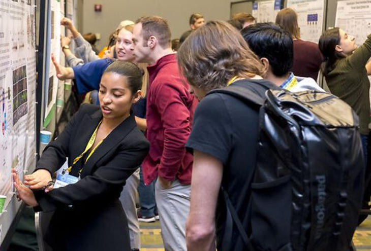
At every conference, everyone focuses on the Keynote and invited talks, but what I look forward to most each year is the “Poster-to-Podium” session—where the conference selects the 10 most innovative posters from hundreds and invites their authors to give brief presentations. These studies lack “endorsements from big-name scholars” or “the prestige of top-tier journals,” yet they often harbor the seeds of “the next major discovery.” They represent the most “unadorned” form of original innovation and serve as a “hotbed” for interdisciplinary research.
4.2.1 Why Does the “Poster Zone” Produce More Dark Horses? — Innovation Unshackled from Paradigms
After spending an entire day exploring the Poster Zone, I noticed a common thread among the research presented here: it defies “traditional disciplinary boundaries,” often combining “cross-frontier” approaches like “cellular sociology + organelle networks” or “spatiotemporal biology + AI.” Since most poster presenters are doctoral students or postdocs, they lack the pressure to “dig deep in a specific field.” This freedom encourages bold experimentation with seemingly unrelated crossovers. Such “unfettered” innovation creates fertile ground for dark horses to emerge.
Take a postdoc team from UCSF, whose poster explored “How Mechanical Forces in the Tumor Microenvironment Regulate Mitochondria-Endoplasmic Reticulum Interactions”—integrating “microenvironmental mechanical forces” from cell sociology with “mitochondria-endoplasmic reticulum interactions” from organelle networks. They discovered that the “high stiffness” of the tumor microenvironment increases contact points between mitochondria and the endoplasmic reticulum, leading to abnormal calcium ion exchange and subsequently promoting cancer cell proliferation. Conversely, reducing environmental stiffness decreases contact points and inhibits proliferation.
I spoke with this postdoc for 20 minutes. He explained that this idea stemmed from “an experimental accident”: originally studying the effects of mechanical forces on cell migration, they unexpectedly observed changes in mitochondrial morphology, which led them to trace alterations in the endoplasmic reticulum. “If I were a senior professor, I might have focused solely on migration and overlooked this ‘accident,'” he said with a smile. “But as a postdoc, I dared to dig deeper into this unexpected finding.” Now, this research has caught the attention of Novartis, which plans to collaborate on developing “drugs targeting mechanosignaling-organelle interactions.”
Another doctoral candidate from the University of Tokyo presented a poster titled “Analyzing Collective Decision-Making in Cell Sociology Using 4D Nucleomics”—applying “4D nucleomics” from “spatiotemporal biology” to study “collective behavior” in “cell sociology.” She discovered that when a group of stem cells decides to differentiate, their chromatin undergoes “synchronized dynamic changes,” akin to a “collective vote.” Disrupting this synchrony causes the stem cells to “hesitate” and fail to differentiate. This finding fills a gap in the “molecular mechanisms of cellular collective behavior” and has been invited for submission to Developmental Cell.
4.2.2 “Poster-to-Podium” Selection Logic: Focus on Breakthroughs, Not Fame
The conference’s criteria for selecting “Poster-to-Podium” presentations are clear: titles and affiliations are irrelevant. The evaluation focuses solely on three dimensions—whether the work “breaks existing paradigms,” “is supported by robust data,” and “can spark new research directions.” Nearly all of this year’s 10 selected posters meet these criteria, with “paradigm-breaking” being particularly prominent.
For instance, a Dutch team challenged the “traditional understanding of organelle interactions”: Previously, it was believed that “mitochondria-endoplasmic reticulum contact sites exist only in the cytoplasm.” However, using super-resolution microscopy, they discovered such contact sites also exist within the nucleus and can regulate chromatin states. This discovery directly redefines the spatial scope of organelle interactions. One EMBO keynote speaker remarked: “Our previous research was confined to the cytoplasm. Now it appears interactions within the nucleus may be more significant—this poster has opened up a new direction for us.”
Another selected team from the US used AI to predict “tunnel nanotube (TNT) formation in cellular sociology,” breaking the paradigm that “TNTs could only be observed experimentally.” Their trained AI model predicts the probability and direction of TNT formation based on cell morphology and gene expression, achieving 89% accuracy—10 times faster than traditional observation methods. An ASCB judge remarked: “This research not only enhances efficiency but, more importantly, enables us to ‘predict in advance’ cellular social behaviors, opening a window for intervention.”
4.2.3 Prediction: The Next “Major Discovery” Lies at the “Crossroads of Research Frontiers”
Based on observations in this year’s poster session, I believe the major breakthroughs in the next 3-5 years will most likely emerge at the “intersections” of the three major research domains mentioned earlier—not through deepening within a single domain, but through cross-domain integration. Specifically, three intersecting directions warrant close attention:
The first is “Cell Sociology × Organelle Networks”: Examples include the previously mentioned “mechanical force regulation of mitochondrial-endoplasmic reticulum interactions” and “how intercellular communication influences organelle interaction networks.” This direction could explain why identical organelles exhibit different functions across distinct cell populations. For instance, mitochondria in cancer cells promote proliferation within tumor populations, whereas they drive metabolism in normal cells.
The second is “spatiotemporal biology × cellular sociology”: Examples include “4D nucleomics analyzing synchronized behavior in cell populations” and “live imaging tracking dynamic changes in cellular social networks.” This direction fills gaps in understanding “spatiotemporal mechanisms of cellular collective behavior,” such as how cells in embryonic development “form tissues precisely on schedule” rather than growing chaotically.
The third is “spatiotemporal biology × organelle networks,” exemplified by “4D imaging of dynamic organelle interactions” and “in situ multi-omics analysis of the molecular basis for organelle interactions.” This direction addresses the “temporal dimension gap in organelle interactions,” such as whether organelle interactions during apoptosis undergo “sudden changes” or “gradual changes,” and identifying critical temporal points.
This year’s “Poster-to-Podium” program already features three studies spanning these three interdisciplinary directions, all attracting significant attention from both academia and industry. This demonstrates that such “cross-disciplinary innovation” has become an industry “consensus” and will be a “mainstream direction” for the next decade.
Table 4-2 Core Innovation Cases from the 2025 Conference “Poster-to-Podium”
| Selected Poster Topic | Cross-disciplinary Direction | Core Breakthrough Points | Data Support / Experimental Results | Industry Impact / Potential Applications | Focused Institutions |
| Mechanical force regulation of the tumor microenvironment Mitochondria-endoplasmic reticulum interactions | Cell Sociology × Organelle Network | First discovery that microenvironmental stiffness influences calcium exchange via contact points, thereby regulating cancer cell proliferation | High-hardness environments increase contact points by 30%, doubling cancer cell proliferation rates; reducing hardness inhibits proliferation by 40% | Developing Anti-Cancer Drugs Targeting Mechanochemical-Organelle Interactions | Novartis, Penn Cancer Center |
| 4D Nucleomics Reveals Synchrony in Stem Cell Population Differentiation | Spatio-temporal biology × cellular sociology | Discovery of “synchronized dynamic chromatin changes” during stem cell differentiation; disruption of synchrony causes differentiation arrest | Highly synchronized stem cell differentiation efficiency reaches 85%, while low synchrony yields only 30% | Optimizing directed stem cell differentiation for regenerative medicine | Stanford Center for Regenerative Medicine, FDA |
| AI predicts formation of tunnel nanotubes (TNTs) | Cell Sociology × AI Technology | Predicting TNTs based on cell morphology/gene expression with 89% accuracy and 10x efficiency improvement | Predict TNT formation timing with <2-hour error margin—10x faster than traditional observation | Intervene early in cellular communication to inhibit cancer metastasis | DeepMind, Pfizer |
| Discovery of mitochondria-endoplasmic reticulum contact points within the cell nucleus | Organelle networks × spatiotemporal biology | Breaking the perception that “contact points exist only in the cytoplasm,” discovering nuclear contact points regulating chromatin | Abnormal nuclear contact sites correlate with chromatin reprogramming in lung cancer cells, occurring in 60% of cases | Developing cancer therapies targeting nuclear contact sites | MIT, AstraZeneca |
4.3 Awards and Funding: Viewing Academia Through a “Venture Capital” Lens — Which Research Tracks Are Capital and Foundations “Betting” On?
Conference award ceremonies and funding announcements serve as industry “capital barometers”—which research wins awards and which directions secure funding reveal foundations’ and capital’s judgments on “future frontiers.” This year’s Cell Bio 2025 awards and funding demonstrate clear “focus”: rather than being dispersed across multiple fields, they concentrate on several core directions. These areas are also highly likely to be the “heavy investment zones” for capital and academic resources over the next decade.
4.3.1 Core Awards: Focus on “Cross-Boundary Innovation” and “Translational Potential”
The three major core awards at this year’s conference—the “ASCB Young Scientist Award,” the “EMBO Molecular Biology Prize,” and the “Cell Bio Translational Research Award”—all share a common thread: the winning research either represents “cross-disciplinary innovation” or possesses “clear translational potential.” None represent “traditional research confined to a single field.”
The “ASCB Early Career Award” went to an assistant professor at Stanford University whose research involves “using AI + in situ multi-omics to decipher cellular social networks in the tumor microenvironment”—spanning the fields of “cellular sociology,” “spatiotemporal biology,” and “AI technology.” Her developed technology simultaneously analyzes “cell location, gene expression, signaling molecules, and organelle status” on a single tumor section. By constructing “cell social network models” through AI, it precisely identifies “core communication nodes within tumors.” This technology has been applied to precision lung cancer treatment, helping physicians determine “which cells are key therapeutic targets” and doubling patients’ progression-free survival.
The EMBO Molecular Biology Prize was awarded to a team from Heidelberg University in Germany for their research on “dynamic analysis of the organelle interactome.” — focusing on the “organelle network” battlefield while breaking the paradigm of “static research.” Using 4D imaging technology, they tracked “dynamic changes in organelle interactions throughout the cell lifecycle,” discovering that “during cellular aging, mitochondrial-endoplasmic reticulum contact points gradually decrease, leading to energy metabolism disorders.” Though fundamental research, this work provides a clear target for “anti-aging drug development” and has secured follow-up funding from the Gates Foundation.
The most direct award, the Cell Bio Translational Research Prize, went to the University of Pennsylvania team for their “artificial mitochondria therapy for mitochondrial genetic diseases”—a cross-disciplinary approach combining synthetic biology and organelle networks. Their engineered “artificial mitochondria” can be delivered intravenously into patient cells to restore impaired mitochondrial function. In mouse studies, this approach achieved a 70% symptom relief rate for mitochondrial disorders and will enter human clinical trials next year. This research has already attracted $120 million in investment from Vertex Pharmaceuticals to advance clinical development.
4.3.2 Funding Directions: Capital and Foundations’ “Betting List”
Beyond awards, the conference announced key funding priorities for 2025-2026. Support from institutions including NIH (National Institutes of Health), EMBO Foundation, Gates Foundation, and Pfizer focuses on four tracks, each explicitly requiring “interdisciplinary collaboration”:
The first track is “AI + Fundamental Cell Biology Research”: NIH is investing $200 million to fund projects “using AI to decipher the mechanisms of cellular networks (e.g., organelle interaction networks, cellular social networks)”, requiring each project to include both “cell biologists + AI algorithm engineers”. For example, a team at MIT received $3 million to “use AI to predict dynamic changes in mitochondrial-endoplasmic reticulum contact points,” aiding in deciphering the mechanisms of neurodegenerative diseases.
The second track is “Cell Sociology and Disease Therapy”: The EMBO Foundation has invested €50 million to fund “disease intervention research based on cellular social networks,” focusing on cancer and autoimmune diseases. For instance, a Cambridge University team received €4 million to study “disrupting tumor cell social networks to inhibit metastasis,” planning to develop “small-molecule drugs that block tunnel nanotube formation.”
The third track is “Synthetic Biology and Organelle Engineering”: The Gates Foundation has invested $100 million to fund “artificial organelle therapies for rare diseases,” particularly mitochondrial disorders and lysosomal storage diseases. The University of Pennsylvania’s “artificial mitochondria” team received $20 million to advance clinical research. Additionally, a Chinese team received $15 million to develop “artificial lysosomes for treating Gaucher disease.”
The fourth track is “spatiotemporal biology technology development”: Pfizer has invested $80 million to fund “high-resolution in vivo imaging technologies” for drug screening. For example, a Stanford University team received $5 million to develop a “live imaging system capable of simultaneously tracking five organelles,” assisting Pfizer in screening “drugs that do not disrupt organelle interactions” to reduce drug toxicity.
4.3.3 The Essence of the Game: Is Capital “Defining” Academic Directions?
Some worry: Could concentrated funding from capital and foundations cause academic research to “follow the money,” losing its autonomy? But discussions at this conference clarified: It’s actually a “two-way selection”—capital bets on promising directions, yet these directions themselves represent academic “consensus”; simultaneously, academic research requires capital support to transition from “laboratory” to “application.”
For instance, funding for “AI + Cell Biology” didn’t emerge from capital’s imagination. It arose because academia recognized that “AI solves problems traditional methods cannot” (like analyzing complex networks). Capital investment then accelerates this direction—developing more precise AI models and more efficient experimental tools. As the NIH funding director stated: “We are not ‘defining’ directions, but ‘accelerating’ those already proven to have potential.”
This “mutual selection” process is bringing academia and industry “closer together”—whereas academic research and capital were once “parallel lines,” they now occupy different segments of the “same line.” Take the University of Pennsylvania’s “artificial mitochondria” research as an example: it began with an academic breakthrough, secured funding, attracted industrial investment, and ultimately advanced to clinical trials, forming a closed loop of “academia-capital-industry.” This closed-loop model will become the “norm” for cell biology development over the next decade.
Table 4-3 Key Funding Tracks and Case Studies for 2025-2026
| Funding Tracks | Primary Funding Institutions | Funding Amount | Core Requirements (Interdisciplinary/Translational) | Representative Funding Cases | Expected Outcomes (3-5 Years) |
| AI + Cell Biology Basic Research | NIH, DeepMind | $230 million (NIH $200 million + DeepMind $30 million) | Requires collaboration between cell biologists and AI engineers, focusing on network mechanism analysis | MIT Team ($3 million): AI prediction of mitochondrial-endoplasmic reticulum contact point dynamics | Develop 5-8 cellular network AI prediction models to decipher 2-3 disease mechanisms |
| Cell Sociology and Disease Therapy | EMBO Foundation, Novartis | €90 million (EMBO €50 million + Novartis €40 million) | Design intervention strategies based on cellular social networks, targeting cancer/autoimmune diseases | Cambridge team (€4 million): Small-molecule drug development targeting TNTs | 3-5 preclinical intervention strategies, with 1-2 advancing to Phase I clinical trials |
| Synthetic biology and organelle engineering | Bill & Melinda Gates Foundation, Vertex Pharmaceuticals | $1.12 billion (Gates $100 million + Vertex $120 million) | Artificial organelles for rare disease treatment require preclinical data validation | Penn team ($20 million): Clinical studies of artificial mitochondria | 2-3 artificial organelles enter clinical trials; 1 approved for mitochondrial genetic disorders |
| Spatio-temporal biology technology development | Pfizer, Zeiss | $85 million (Pfizer $80 million + Zeiss $5 million) | Developing high-resolution in vivo imaging technology for drug screening | Stanford team ($5 million): 5 synchronized organelle tracking systems | 1-2 imaging technologies for pharmaceutical screening, reducing drug toxicity rates by 30% |
V. Conclusion: Bringing the “Spark” Back to Philadelphia—Action Guide for Attendees and “Virtual Participants” of Cell Bio 2025, the Flagship Bio US Conference
Returning from the conference, my suitcase held few souvenirs—just three sheets of paper: a hand-drawn “core venue map,” an agenda marked with “must-attend presentations,” and contact details from a PhD student I met in the poster area These three sheets of paper represent the true “sparks” worth carrying home from this conference: not scattered knowledge points, but “actionable plans” and “sustainable connection opportunities.” Below are practical guides tailored for on-site attendees and online participants, followed by a glimpse into what we might be discussing in 2026 as a result of this conference.
5.1 For You “On-Site”: Three “Most Efficient” Agenda Browsing Paths
Last year, during my first Cell Bio conference, I made a rookie mistake: I didn’t plan my route in advance. With the mindset of “wanting to explore every venue,” I ended up exhausted and unproductive—missing key presentations and failing to engage deeply with exhibitors. This year, I did my homework and mapped out three paths based on my “core needs,” focusing on one area each day—doubling my efficiency. If you’re still at the Philadelphia venue, here are three paths you can follow:
5.1.1 Path One: “Technology-Driven” — Focus on Tools and X-Factors, Hands-On Learning
Ideal for: Lab technical leads, grad students looking to upgrade experimental methods, professionals interested in AI/synthetic biology
Core Logic: Master “immediately applicable tools” first, then explore “future-driving technologies,” transforming “conference attendance” into “skills training”
Sample Day 2 Schedule:
- 09:00-10:30: Hands-on workshop at the tool exhibition (focus on 10x Genomics’ Xenium Flex HD and Nikon’s in situ cell interaction microscope) — Last year I only observed; this year I pre-booked a “1-on-1 demo,” mastering basic spatial transcriptomics analysis in 20 minutes. Now I can train my lab team upon return.
- 11:00-12:30: “AI + Cell Biology” Special Forum (listen to DeepMind team present AlphaCell real-world applications) — Bring a USB drive to collect AI analysis starter scripts on-site, saving time searching for resources;
- 14:00-15:30: Synthetic Biology Tools Track (Focus on practical details of “Chassis Cell Libraries” and “Artificial Organelle Design”) — The University of Pennsylvania team will distribute the Synthetic Cell Experiment Manual, which includes pitfall avoidance guides (e.g., how to prevent cytotoxicity during artificial mitochondrial delivery);
- 16:00-17:30: “Technical Tools” Zone in the Poster Area (Look for booths labeled “Trial Available,” such as the OrganellePlex software team. Fill out an application on-site for a free 3-month trial).
5.1.2 Pathway Two: “Disease-Driven Approach” — Anchoring Clinical Needs to Identify Translation Opportunities
Suitable for: Pharmaceutical R&D personnel, clinicians, researchers focused on disease mechanisms
Core Logic: Backtrack from “clinical pain points” to identify “research directions,” prioritizing engagement with teams possessing translation potential
Specific Schedule (Example: Day 3 of the Conference):
- 08:30-10:00: “Sociology of Cancer Cells” Breakout Session (Hear the Harvard team discuss “Tumor Microenvironment Cell Interactions and Drug Resistance,” featuring live comparisons of clinical sample data, such as variations in TNT distribution among different patients);
- 10:30-12:00: “Neurodegeneration and Organelle Networks” Roundtable (Cambridge’s Haass team will share latest Alzheimer’s disease mechanisms; 15-minute Q&A session follows—prepare clinical challenges like “methods for detecting organelle fragments in patient cerebrospinal fluid”);
- 13:30-15:00: “Research-Industry Matchmaking Session” (Pfizer and Novartis booths feature “Need Walls” listing urgent technical challenges, e.g., “Rapid screening of drugs that preserve organelle interactions.” If your research aligns, leave contact details—you’ll likely receive collaboration invitations);
- 15:30-17:00: Tour of the “Clinical Translation Center” at Children’s Hospital of Philadelphia (pre-register via the conference website to observe organoid applications in clinical settings, such as predicting pediatric drug responses using intestinal organoids).
5.1.3 Path Three: “Pure Basic Science Approach” — Deep Dive into Three Major Research Frontiers, Following Leading Experts’ Thinking
Suitable for: Basic research scholars, doctoral students (seeking research directions), students interested in disciplinary frontiers
Core Logic: Focus on “fundamental mechanism reports” across three major research domains, prioritizing understanding “pioneers’ thought processes” over merely absorbing “conclusions.”
Sample Schedule (Day 1):
- 09:00-10:30: EMBO Basic Research Session (Listen to Venki Ramakrishnan discuss “Ribosomal Quality Control Mechanisms”; note “unresolved questions” he mentions, such as “links between ribosomal assembly errors and rare diseases”—potential future research entry points);
- 11:00-12:30: “Cellular Sociology and Collective Intelligence” session (MIT’s Maria Dominguez team will share approaches to constructing “tumor cell social networks,” e.g., “how to identify key signaling molecules from spatial transcriptomics data”; clearly document analytical steps for potential reuse in your own research);
- 14:00-15:30: “Spatio-Temporal Biology: 4D Nucleomics” Session (Stanford team will present raw data from chromatin dynamics tracking. Pay attention to their “data analysis methods,” such as background noise exclusion techniques);
- 16:00-18:00: “Big Names Tea Break” (Daily at 4 PM in the main venue lounge, featuring keynote speakers on duty. Last year I didn’t dare attend, but this year I mustered the courage to ask Professor Bertozzi about “the next frontier in glycosylation research.” She recommended three pivotal papers—a gain far greater than any lecture).
Table 5-1 Comparison of Three On-Site Participation Pathways
| Path Type | Suitable Audience | Core Sessions (Including Time/Location) | Essential Items/Tools | Pitfall Reminders |
| Technology-Driven | Lab technical leads, graduate students seeking advancement methods | 09:00-10:30 Tools Exhibition Area (10x / Nikon Booth); 14:00-15:30 Synthetic Biology Tools Breakout Session | USB drive (for scripts/manuals), laptop (for on-site software testing) | Don’t just watch demos—hands-on practice is essential. For example, adjust microscope settings yourself |
| Disease-Driven | Pharmaceutical R&D, clinicians, translational researchers | 10:30-12:00 Neurodegenerative Roundtable (Hall 3); 13:30-15:00 Industry Matchmaking Session (Exhibition Area C) | Clinical case notes (with patient data for easy questioning), business cards (print 50 extra) | When meeting pharmaceutical companies, don’t just say “My research is impressive.” Instead, explain “How it addresses your specific pain points.” |
| Pure Basic Science Researchers | Basic scientists, PhD students (seeking research topics) | 09:00-10:30 EMBO Session (Hall 1); 16:00-18:00 Senior Scientist Networking Break (Main Venue Lounge) | Research notebook (list your research challenges), literature list (for recommendations) | Avoid broad questions (e.g., “How do I choose a research direction?”). Be specific (e.g., “What do you see as the key limitations of XX experimental method?”) |
5.2 For Online Attendees: Three Must-See Debates and Must-Hear Talks
Many online attendees have shared with me: “It feels disconnected through the screen—I’m unsure what to focus on.” The key to online participation is “focusing on core content + finding interactive channels.” The conference website and ASCB/EMBO communities offer many “online-exclusive resources”—debate replays, presentation slides, high-resolution poster images. If you know where to look, your takeaways can rival an in-person experience. Here’s my curated “must-see list”:
5.2.1 Must-Watch Debate 1: “Will AI Reshape the Research Paradigm in Cell Biology?” (November 20, 15:00-16:30; replay link available in the “Hot Topics” section of the conference website)
This debate was fiercely charged! On one side, a DeepMind AI engineer argued that “within five years, AI will dominate hypothesis generation, with wet labs solely responsible for validation.” Opposing him, a senior cell biologist from the University of Pennsylvania countered that “AI can only predict based on existing data and cannot discover unknown phenomena.” One particularly intriguing detail emerged: The engineer presented AI-predicted “undiscovered mitochondrial-endoplasmic reticulum contact proteins,” while the biologist immediately challenged, “How would you design experiments to validate these proteins’ functions?” Both even agreed to collaborate on verification experiments post-debate. When watching the replay online, focus on “both sides’ arguments and experimental examples”—not just conclusions. These cases will help you gauge AI’s applicability within your own research.
5.2.2 Must-Watch Debate 2: “Basic Research vs. Translational Research: Which Should Get Priority Funding?” (November 21, 10:00-11:30, replay with real-time chat discussion)
Debaters include the EMBO Foundation’s funding director (advocating basic research priority) and Pfizer’s R&D director (supporting translational research priority). The EMBO side presents compelling arguments: “Without breakthroughs in basic research, translational work is like cooking without ingredients—for instance, without discovering the lysosome-endoplasmic reticulum interaction mechanism, targeted drugs for Alzheimer’s disease wouldn’t exist.” Pfizer counters: “Focusing solely on basic research leaves many findings confined to papers, unable to benefit patients.” The online format allows viewing “industry polls” via real-time comments. During my viewing, 60% supported “funding basic and translational research at a 7:3 ratio”—a valuable benchmark for both grant applications and research planning.
5.2.3 Must-See Presentations: Three Paradigm-Shifting Keynotes (Free PPT downloads available on ASCB website, featuring speaker-annotated “key pages”)
- Carolyn Bertozzi (ASCB): “From 0 to 1 in Glycosylation-Targeted Therapies” — Focus on the “timeline from mechanism discovery to clinical application” (slides 12-15), which outlines key milestones and pitfalls at each stage (e.g., addressing antibody immunogenicity in Phase I trials). Essential viewing for those pursuing translational research;
- Christian Haass (EMBO): “Disrupted Organelle Interactions and Neurodegenerative Diseases” — Slides 8-10 feature “cryo-EM structures of lysosome-endoplasmic reticulum contact sites” with annotated binding sites for mutant proteins. Directly applicable for structural biology or disease mechanism research.
- Li Feifei Team (Spatio-Temporal Biology Session): “AI + Live Imaging: New Tools for Studying Cell Dynamics” — The final slide includes a “free trial application link for AI imaging tools” and FAQs (e.g., noise handling in imaging data). Online attendees can apply directly to avoid seeking alternative channels.
Table 5-2: Must-See/Must-Hear List for Online Attendees
| Content Type | Specific Topic | Time / Access Method | Core Value (Online Exclusive) | Viewing/Learning Recommendations |
| Hot Debate | Will AI Reshape the Paradigm of Cell Biology Research? | November 20 Replay (Conference Website > “Hot Topics”) | Review experimental cases from both sides to assess AI applicability boundaries; use real-time polls to gauge industry consensus | While watching, note “AI applications you can try in your own research,” such as “using AI to predict cell migration trajectories” |
| Hot Debate | Funding Priorities for Basic vs. Translational Research | November 21 Replay (with live comments) | Understand funding preferences; reference the “7:3” resource allocation ratio | Align with your research to plan “integration points between basic and translational research,” such as “conducting mechanism studies before designing translational approaches” |
| Keynote Presentation | Bertozzi: From 0 to 1 in Glycosylation-Targeted Therapy | Download PPT from ASCB official website (with key points highlighted) | Reference the translation timeline; obtain pitfall avoidance guidelines for clinical stages | Focus on the “Clinical Data Comparison” page to align your research with clinical needs |
| Keynote Presentation | Haass: Organelle Interactions and Neurodegenerative Diseases | EMBO Official Website Download PPT (Includes Structural Data) | Direct reuse of cryo-EM structures; identify research entry points for mutant proteins | Save the “contact point protein list” for database comparison later to see if it includes proteins relevant to your research |
| Online Interaction | Poster-to-Podium HD Replay + Q&A | Large Community Groups (WeChat / LinkedIn groups) | Ask authors questions (leave comments or schedule 1-on-1 online discussions) | Select 3 interdisciplinary studies (e.g., Cell Sociology × Organelle Networks) to explore experimental design insights |
5.3 Final Prediction: Cell Bio 2025 Casts a “Long Shadow”—What Will We Be Discussing in 2026 Because of This Conference?
At closing, the ASCB President stated: “A great conference isn’t one where attendees feel ‘fulfilled’ at the end, but one that sparks ‘new discussions and research’ a year later.” Based on this conference’s technological signals, industry movements, and funding trends, I boldly predict: By 2026, three “hot topics” will dominate cell biology—and their “seeds” were planted at this Philadelphia conference.
5.3.1 Hot Topic 1: The Initial Implementation of “AI Cell Design Platforms” Sparks Discussions on “Human-Machine Collaborative Research”
At this conference, the “AI Cell Design Platform” developed by DeepMind and MIT is still in the demonstration phase, yet five pharmaceutical companies and ten laboratories have already pre-booked trials. By 2026, we will likely see the first wave of research on “AI-designed cells,” such as AI-engineered “tumor-targeting artificial islet cells,” ” stem cells optimized for organelle interactions,” and potentially even “AI-predicted novel cellular functions” validated through experimentation.
This will spark discussions on “human-machine collaboration”: questions like “Should AI-designed experimental protocols require human review?” and “How do we balance AI efficiency with human innovation?” I can even envision a dedicated “AI-Designed Cells” session at the 2026 Cell Biology Conference, showcasing diverse cell designs created by different teams using the same platform. This would highlight the contrast between “AI’s diversity” and “human-driven specificity”—a debate poised to fundamentally reshape how we conduct research.
5.3.2 Hot Topic #2: Dramatic Cost Reduction in Cell Therapy Fuels “Universal Access” Debate
Novartis revealed at this conference that CAR-T production using “standardized chassis cells” has dropped to $150,000, with plans to launch a lung cancer CAR-T therapy under $200,000 by 2026. — While still substantial, this represents a significant reduction from current million-dollar price tags, making it accessible to more patients and paving the way for cell therapy to transition from treating rare diseases to common ones.
By 2026, we will witness intensified discussions on “making cell therapy accessible to all,” including: “how to further reduce costs (e.g., through synthetic biology-optimized production)”, “whether health insurance should cover cell therapy”, and “how to standardize cell therapy protocols across regions”. Competition may even emerge around “universal cell therapies,” with companies like Pfizer and Bayer launching CAR-T treatments targeting various cancers, competing on “efficacy” and “price.” Ultimately, patients will benefit from this competition, transforming cell therapy from a “niche treatment” into a “mainstream option.”
5.3.3 Hot Topic Three: Explosive Cross-Domain Research Results Reshape the “Disciplinary Framework of Cell Biology”
Both the conference poster sessions and funding directions indicate that cross-disciplinary research integrating “cell sociology × organelle networks” and “spatiotemporal biology × AI” has become a prevailing trend. By 2026, these areas are likely to yield a wave of top-tier journal publications—such as “4D Nucleomics Reveals Dynamic Changes in Tumor Cell Social Networks” and “AI Predicts Diseases Caused by Mitochondrial Interaction Dysregulation.” We may even witness the emergence of new cross-disciplinary theories, like “Spatio-Temporal Dynamics of Cell Populations Determine Mitochondrial Interaction Patterns.”
These findings will gradually reshape the “disciplinary framework” of cell biology—whereas we previously categorized fields by “molecules/organelles/cells,” future classifications may shift toward “networks/systems/dynamics.” New directions like “cellular network biology” and “spatiotemporal systems biology” will emerge. By 2026, when discussing cell biology, we may no longer ask, “Which organelle do you study?” but rather, “Which cellular network dynamics do you investigate?” This reframing will usher in a “new golden decade” for the entire discipline.
Table 5-3 Predicted Hot Topics in Cell Biology for 2026
| Hot Topics in 2026 | Signals from the 2025 Conference | Potential Key Advances in 2026 | Impact on the Industry |
| AI-powered cell design platforms become operational | DeepMind-MIT Demonstrates AI-Designed Artificial Islet Cells; 5 Pharma Companies Book Trial Usage | First Nature/Cell paper on “AI-designed cells for disease treatment”; 2-3 AI-designed cells advance to preclinical stage | Redefining research paradigms, propelling “human-machine collaboration” into mainstream practice |
| Democratizing Cell Therapy | Novartis reduces CAR-T cost to $150,000 using chassis cells; plans 2026 launch for lung cancer CAR-T | Cell therapies priced under $200,000 enter market; universal CAR-T targets 3-5 cancers; some countries include cell therapies in healthcare coverage | Cell therapy expands from rare to common diseases, broadening patient access |
| Explosive growth in cross-warzone research outcomes | 30% of poster presentations feature cross-disciplinary research; NIH funds “Cell Sociology × Organelle Network” project | Number of cross-disciplinary top-tier journal papers doubles; emergence of new “spatiotemporal biology of cellular networks” research direction; 1-2 cross-disciplinary theories proposed | Redefining disciplinary frameworks to advance the field from “single-point research” to “systemic research” |